; ?>)
×

; ?>)
| PARAMETER | SPECIFICATION |
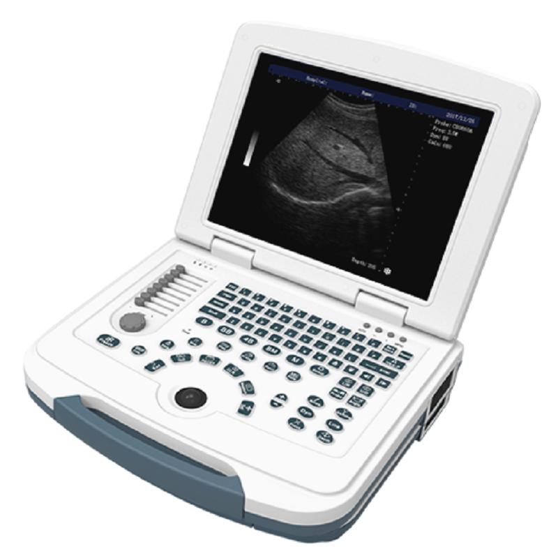
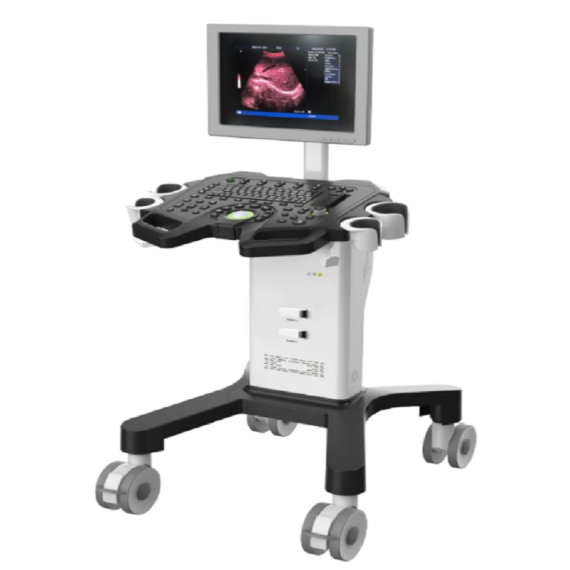
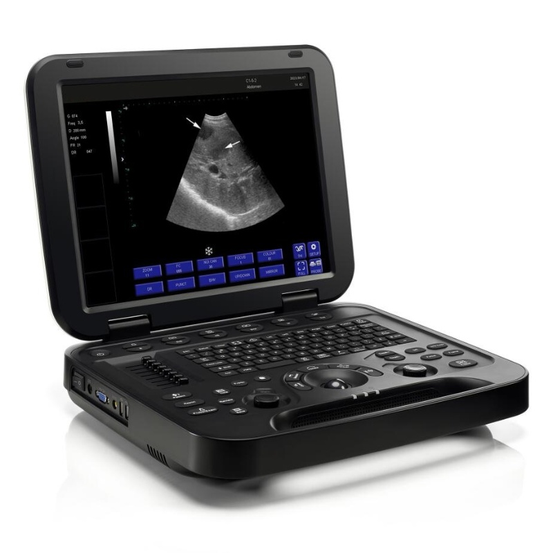
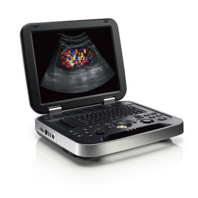
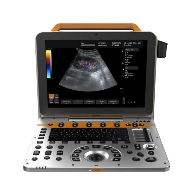
| Portable type all digital color Doppler ultrasonic mainframe | |
| Applications | Abdomen, obstetrics, gynecology, heart, urinary system, small organs, superficial, blood vessels, pediatrics, newborns, musculoskeletal |
| Probes | Convex probe, Tran-vaginal probe, Linear probe, Micro-convex probe, Cardiac probe,4D Volume probe |
| Carotid artery intima measurement thickness(IMT) | |
| Automatic spectral envelope measurement | |
| Full digital transmission and reception of beam synthesizer | |
| Color Doppler imaging (C) | |
| Pulse Doppler Imaging (PW) | |
| Coherent Contrast imaging(CCI) | |
| Continuous wave Doppler imaging(CW) | |
| B/C/D Real-time three synchronous imaging | |
| Power Doppler imaging(PDI) | |
| Direct power Doppler imaging(DPDI) | |
| M mode imaging | |
| Anatomic M mode imaging | |
| Color Doppler M mode imaging | |
| Elastography | |
| Tissue Doppler imaging(TDI) | |
| Strain rate imaging (SRI) | |
| Tissue harmonic imaging(THI) | |
| Fusion harmonic imaging(FHI) | |
| Speckle Reduce imaging(SRI) | |
| Panoramic imaging | |
| Deflection imaging | |
| Trapezoidal imaging | |
| Adaptive velocity optimization | |
| Free hand 3D | |
| Real time 3D imaging(3D/4D) | |
| DICOM3.0 | |
| Monitor | 15 inch,high definition ultrasonic display |
| Physical clipboard | save the image on the left side of the screen, which can be directly saved or deleted. |
| The system has the function of on-the-spot upgrade | |
| Presupposition | for different inspection of the viscera, preset the inspection conditions for the best image, reduce the adjustment of the operation, and the commonly used external adjustment and combination regulation. |
| Probe interface | 2 |
| System languages | Chinese/English/ Spanish/ Portuguese/ French/ Russian/ Arabic/
Vietnamese/ Indonesian/ German |
| Depth | ≥360mm |
| Extended imaging | |
| Probes | |
| Convex probe | Fundamental Frequency:
2.0MHz/2.3MHz/2.5MHz/3.0MHz/3.5MHz/4.0MHz/4.6MHz/5.0MHz/5.4MHz |
| Harmonic Frequency:
4.0MHz/4.6MHz/5.0MHz |
|
| Linear probe | Fundamental Frequency:
4.0MHz/4.6MHz/5.0MHz/6.0MHz/7.0MHz/8.0MHz/9.2MHz/10.0MHz/12.0MHz/13.3MHz |
| Harmonic Frequency:
8.0MHz/9.2MHz/10.0MHz |
|
| Trans-vaginal probe | Fundamental Frequency:
3.0MHz/3.5MHz/4.0MHz/5.0MHz/5.4MHz/6.0MHz/7.0MHz/8.0MHz/10.0MHz |
| Harmonic Frequency:
6.0MHz/7.0MHz/8.0MHz |
|
| Micro-convex probe | Fundamental Frequency:
3.0MHz/3.5MHz/4.0MHz/5.0MHz/5.4MHz/6.0MHz/7.0MHz/8.0MHz |
| Harmonic Frequency:
6.0MHz/7.0MHz/8.0MHz |
|
| Cardiac probe | Fundamental Frequency:
1.7MHz/1.9MHz/2.1MHz/2.5MHz/3.0MHz/3.4MHz/3.8MHz/4.2MHz/5.0MHz |
| Harmonic Frequency:
3.4MHz/3.8MHz/4.2MHz |
|
| 4D volume probe | Fundamental Frequency:
2.0MHz/2.5MHz/3.0MHz/3.3MHz/3.7MHz/4.0MHz/5.0MHz/6.0MHz |
| Harmonic Frequency:
4.0MHz/5.0MHz/6.0MHz |
|
| 2D Imaging Mode | |
| Gain | 0-100,Step 2 adjustable |
| TGC | 8 segment adjustable |
| Maximum focus point | ≥7, which can be moved throughout the whole process |
| Speckle reduction | 0-5,5 level |
| Space Synthesis | 0-2,2 level (Liner probe: 3 level,cardiac probe:0) |
| Dynamic | 30-180,35 level,step 5 adjustable |
| Line density | low、middle、high,3 level |
| Frame correlation | 0-4, 4 level |
| Noise reduction | 0-5, 5 level |
| Edge Enhancement | 0-5, 5 level |
| Sound power | 2-10, 9 level |
| Grey scale | 0-67, 67 level |
| Grey scale | 0-67, 67 level |
| Image style | Soft-Comparison,2 level |
| The screen has real-time display of voice power, probe frequency, dynamic range, pseudo color, gray scale and other 11 parameters can be adjusted | |
| Color Doppler Imaging Mode | |
| Blood gain | 0-100,Step 2 |
| Parameter display | Velocity、Variance |
| B-Restrain
(B/W restrain) |
0-7, 7 level |
| Speed through | 0-8, 8 levels |
| Sampling number | 6-24, 7 level |
| Blood flow preferred | 0-8, 8 level |
| Filtering | 1-6, 6 level |
| Sound power | 2-6, 4 level |
| Noise reduction | 0-4, 4 level |
| Smooth treatment | 0-4, 4 level |
| Frame correlation | 0-6, 6 level |
| Chromatography
(Blood flow graph) |
0-37, 37 level |
| Line density | Low-Middle-High,3 level |
| Frequency | 4 level adjustable |
| Velocity | Minimum 0.4K,Maximum 40.5K |
| Convex probe | 0.4K-4.3K-38.5K |
| Linear probe | 0.4K-14.7K-39.0K |
| Trans-vaginal probe | 0.4K-7.8K-39.7K |
| Volume probe | 0.4K-4.2K-34.8K |
| Micro-convex probe | 0.4K-10.3K-40.5K |
| cardiac probe | 0.4K-7.8K-39.7K |
| PS:The frequency of the probe changes and the frequency value changes | |
| PS:Frame rate changes with speed | |
| Pulse Wave Doppler (PW) | |
| Gain | 0-100,Step 2 |
| Spectrum envelope function | real time automatic spectrum envelope, manual spectrum envelope, and other modes. The system automatically analyses and displays various data such as PSV, EDV, RI, PI, S/D, ACC, HR and so on. Can wake up or close |
| Sample volume | 0.5mm~30mm |
| Blood angel | -75—75 degree,Step 5 |
| False color | 0-67,67 level |
| Dynamic range | 20-40,4 level |
| Filter | 0-9, 9 level |
| Smooth treatment | 1-4,4 level |
| Sound power | 2-5,4 level |
| Volume | 0-100, 10 level, Step 10 |
| Audio filtering | 0-4,4 level |
| Base line | -1.0~1.0 |
| Grey map | 0-67, 67 level |
| Scan velocity | 100-500, 6 level |
| PRF | Minimum 0.5K,Maximum 87.5K |
| Convex probe | 0.5K-4.3K-63.3K |
| Linear probe | 0.5K-14.5K-78.4K |
| Trans-vaginal probe | 0.5K-8.1K-78.4K |
| Volume probe | 0.5K-4.2K-53.8K |
| Micro-convex probe | 0.5K-10.3K-81.1K |
| Cardiac probe | 0.5K-4.3K-87.5K |
| Frequency | 4 level |
| PS:The frequency of the probe changes and the PRF value changes | |
| PS:The frequency of the probe changes and the frequency value changes | |
| Continuous Wave Doppler(CW) | |
| Support probe | Cardiac probe |
| Adjustment of B mode parameters is switchable | |
| Gain | 0-100,Step 2 |
| Sampling line position is adjustable | |
| PRF | 0.9K~36.1K |
| Baseline | -1.0~1.0 |
| Blood angel | -75~75 degree |
| Grey map | 0-67 |
| Scan velocity | 100-300 |
| False color | 0-67 |
| Dynamic range | 20-40 |
| Filtering | 0-9,9 level |
| Smooth treatment | 1-4 |
| Frequency | 2.0MHz/2.3MHz/2.5MHz/3.0MHz,4 level adjustable |
| Sound power | 2-5 |
| Volume | 0-100 |
| Audio Filtering | 0-4 |
| Anatomical M imaging | |
| Support probe | Convex probe, Linear probe,Cardiac probe |
| Adjustment of B mode parameters is switchable | |
| Gain | 0-100,Step 2 |
| M Sampling line angel is adjustable | |
| M Sampling line length is adjustable | |
| Sampling line | 3, Can be displayed or hidden separately |
| Blood flow M model(MC) | |
| Adjustment of B mode parameters is switchable | |
| Gain | 0-100,Step2 |
| MC Sampling line angel is adjustable | |
| MC Sampling line length is adjustable | |
| Frequency | 4 level |
| Sampling number | 6-24 |
| Speed through | 0-8, 8 level |
| Scan velocity | 150-500 |
| Frame correlation | 0-6, 6 level |
| Filtering | 1-6, 6 level |
| Blood flow preferred | 0-8, 8 level |
| Smooth treatment | 0-4, 4 level |
| Map | 0-37, 37 level |
| Elastography | |
| Adjustment of B mode parameters is switchable | |
| Gain | 0-100,Step 2 |
| B/E,Double real-time display on the same screen | |
| Probe displacement curve display | Up/Down |
| Pressure indicator bar display | |
| Frequency | 8-9 level,Adjustable;According to the probe display |
| Noise reduction | 0-2, 2 level |
| Frame correlation | 0-3, 3 level |
| Comparison | 0-13, 13 level |
| False color | 0-3,3 level |
| Don’t support cardiac probe | |
| Tissue Doppler imaging(TDI) | |
| Support probe | Cardiac probe |
| Adjustment of B mode parameters is switchable | |
| Gain | 0-100,step 2 |
| ROI area adjustable | |
| Sampling number | 6-24 |
| Velocity | 0.4K-8.0K |
| Frame correlation | 0-6,6 levels |
| Tissue preferred | 0-7, 7 level |
| Frequency | 2.0MHz/2.3MHz/2.5MHz/3.0MHz |
| Support color reversal | |
| Strain rate imaging | |
| Support probe | Cardiac probe |
| Adjustment of B mode parameters is switchable | |
| ROI area adjustable | |
| Gain | 0-100,Step 2 |
| Sampling number | 6-24,6 level |
| Axial average | 1-4, 4 level |
| Velocity | 0.4K-8K |
| Frame correlation | 0-6, 6 level |
| Tissue optimization | 0-7, 7 level |
| Panoramic Imaging | |
| Support probe | Linear probe |
| Speckle Reduction | 0-5, 5 level |
| Deflection Imaging | |
| Support probe | Linear probe |
| Adjustment of B mode parameters is switchable | |
| Deflection angel | 8 level |
| Speckle Reduction | 0-5, 5 level |
| Dynamic rate | 30-180,Step 5 |
| Line density | low-middle-high, 3 level |
| Frame Correlation | 0-4, 4 level |
| False color | 0-67, 67 level |
| Image style | Soft-Comparison, 2 level |
| Noise reduction | 0-5, 5 level |
| Edge Enhancement | 0-5,5 level |
| Sound power | 2-10,8 level |
| Grey map | 0-67,67 level |
| Trapezoidal Imaging | |
| Probe support | linear probe |
| Adjustment of B mode parameters is switchable | |
| Deflection angel | 8 level |
| Speckle reduction | 0-5, 5 level |
| Dynamic rate | 30-180, Step 5 |
| Line density | low-middle-high, 3 level |
| Frame Correlation | 0-4, 4 level |
| False color | 0-67, 67 level |
| Image style | Soft-Comparison,2 level |
| Noise reduction | 0-5, 5 level |
| Edge Enhancement | 0-5,5 level |
| Sound power | 2-10,8 level |
| Grey map | 0-67,67 level |
| Space Synthesis | 0-2, 2 level |
| Freehand 3D Imaging | |
| Support probe | convex probe, linear probe |
| Display model | 4 pictures |
| Image Rotation X/Y/Z Axis | |
| Multi-slice Visibility | |
| Real-time 4D Imaging | |
| Support probe | 4D volume probe |
| Adjustment of B mode parameters is switchable | |
| Gain | 0-100,Step 2 |
| Display model | one image、two images、four images |
| Image Rotation | X/Y/Z Axis |
| Multi-slice Visibility | |
| Light&Shade inversion | |
| Smooth | 0-4, 4 level |
| Threshold level | 0-129, Step 3 |
| Transparency | 1-509,Step 10 |
| 4 Kinds Render type | Surface、maximum、minimum、perspective |
| Extended Imaging | |
| Gain | 0-100,Step 2 |
| TGC | 8 segment adjustable |
| Maximum focus point | ≥7, which can be moved throughout the whole process. |
| Speckle reduction | 0-5,5 level |
| Space compound | 0-2,2 level(Linear probe: 3 level,don’t support cardiac probe) |
| Dynamic range | 30-180,35 level,Step 5 |
| Line density | Low、Middle、High,3 level |
| Frame correlation | 0-4, 4 level |
| Noise reduction | 0-5, 5 level |
| Edge enhancement | 0-5, 5 level |
| Sound power | 2-10, 9 level |
| Grey map | 0-67, 67 level |
| False color | 0-67,67 level |
| Image style | Soft-Comparison,2 level |
| Extended level | Maximum 72 level |
| Convex probe | 9 level |
| Trans-vaginal probe | 72 level |
| Micro-convex probe | 29 level |
| Cardiac probe | 40 level |
| 4D Volume probe | 17 level |
| PS:The screen has real-time display of voice power, probe frequency, dynamic range, pseudo color, gray scale and other 11 parameters can be adjusted | |
| PS: When the probe scan range reaches the maximum, the space synthesized is 0. | |
| Measurement and analysis function | |
| General measurement | Distance, area, ellipse, cross line, angle, distance ratio, volume, Volume (ellipse), area ratio, diameter, joint angle |
| Cardiac | Automatic spectrum envelope、LV、Main Pulmonary artery diameter、RVEDd、RVEDs、LVM、LAV、HR、MVF、AO、AR、LVOT、TVF、Pulmonic valve、Pulmonary vein、RV、Doppler fetal heart sound、LVET、LVM、LVMI、AV |
| Vascular | carotid intima (IMT), length stenosis ratio, area stenosis ratio, IMT (back wall), IMT (front wall) |
| OB | Fetal routine、AFI、TW、GS、CRl、OFD、HL、ulna、NT、Fibula、Nbonel、Radial、Tibia |
| GYN | uterus、cervix、corpus uteri/cervix uterus、left ovarian vein、right ovarian vein、dominant follicle、intima thickness |
| Urology | prostate、residual urine、left kidney、right kidney、left suprarenal vein、right suprarenal、left testis、right testis、left seminal vesicle、right seminal vesicle |
| Abdomen | liver、CHD、partal vein diameter、cholecyst、CBD、pancreas、spleen、Internal diameter of abdominal aorta、kidney |
| Small parts | Thyroid |
| Software package:Measurement package、Software package、Medical records management software package | |
| Graphic and text management system | |
| Host build in 2 hard disk (SSD 120+1T),Start fast and stable | |
| Movie playback | ≥1200 frames |
| Internal file information management system: can record patient number, name, check number, check date and so on, and can be searched and managed by numbering, checking number, name and so on. | |
| Type of report is 16 | |
| One key fast report graphic and text management | |
| Interface | |
| USB interface | 2 |
| LAN interface | 1 |
| HDMI interface | 1 |
| AUDIO interface | 1 |
| MIC interface | 1 |
| S-VIDEO interface | 1 |
| Configuration | |
| Portable type full digital color Doppler ultrasound diagnostic system | |
| Optional Probes | Convex array probe
Linear probe Trans-vaginal probe Cardiac probe 4D volume probe |
| ≥13 quick adjusting knobs | |