
A patient may present with persistent cough, unknown source of pain, reduced mobility or even no symptoms at all. Even a full physical examination will not give the complete picture and in many cases, a medical image will supply the crucial evidence of any structural changes.
Earlier identification of abnormalities does not mean that an X-ray machine makes a diagnosis. Instead, the equipment produces clinical images, which are interpreted by a doctor together with the patient’s symptoms, medical history, physical examination findings, and results from other tests. Timely access to clear images can help the clinical team identify suspicious changes earlier and determine whether further clinical evaluation is needed.
ARI is a Chinese medical equipment manufacturer and solution provider with more than two decades of experience in hospital projects. The company combines research, manufacturing, production, and sales, with three production bases covering more than 50,000 square meters. Its factories work under ISO 9001 and ISO 13485 quality management systems. Equipment has been supplied to hospitals, clinics, and healthcare facilities in more than 100 countries, while project support covers installation guidance, operation training, maintenance training, and related technical work.
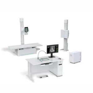
For hospitals that need both routine digital radiography and dynamic fluoroscopy, the ARI AR-7900H High-Frequency Digital Dynamic Radiography System is worth considering. It combines two imaging functions in one system and is designed for radiology departments, medical centers, diagnostic centers, health examination centers, orthopedic departments, and trauma care.
Why Does Earlier Clinical Evaluation Depend on Timely Imaging?
The first image is critical when the patient’s symptoms are mild. It can determine whether the physician will simply monitor the patient or schedule another study, or begin treatment. The value of digital radiography is that it can provide meaningful images to the clinician in a timely manner without adding unnecessary delays to the clinic’s workflow.
Turning Symptoms Into Visible Clinical Evidence
Chest pain, breathing problems, back pain, abdominal discomfort, or restricted joint movement can have many causes. A doctor cannot always separate them through symptoms alone. Digital radiography can show bone alignment, chest structures, joint conditions, and other changes that may explain the patient’s complaint.
This is especially useful when a visible abnormality is still small or when the symptoms do not match the physical examination. The image does not provide every answer, but it gives the doctor a firmer starting point for deciding what should happen next.
Moving From Exposure to Review Faster
Film-based workflows are labor-intensive as they require processing, handling and storage of physical films. In contrast, a digital system captures the acquired radiographic image which is then sent directly to the workstation where the operator can check the positioning of markers and change the viewing window to better evaluate structures of interest. The operator can also add relevant measurements and complete the record for subsequent review.
The difference may be only a few minutes for one patient. Across a busy radiology department, those minutes add up. Faster image access also matters in trauma cases, where the clinical team may be waiting for information before making the next decision.
Where Do Conventional Workflows Delay Clinical Evaluation?
Many imaging delays are not caused by the exposure itself. They come from equipment changes, difficult positioning, repeated examinations, or image files that do not move smoothly through the hospital.
Separate Systems Create Extra Steps
A hospital may use one system for routine radiography and another for fluoroscopy. This means separate room planning, separate operator training, more maintenance work, and patient movement between machines.
A combined system can reduce some of those steps. The AR-7900H provides digital radiography and digital fluoroscopy on one platform, allowing the department to handle static images and real-time examinations without relying on two completely separate systems.
Difficult Positioning Can Cause Repeat Work
Not all patients are able to stand straight and pose in a normal fashion. Some patients are of advanced age, have experienced trauma, or suffer from limited mobility. The positioning of the equipment in relation to the patient and the ability to move the equipment to the required angle or position on the table can be a challenge. It can take a lot of time to reposition a patient and in some cases even an additional exposure is required.
The system uses a four-way floating table with electromagnetic braking. The tube is mounted to move vertically as well as to rotate from -90° to 90° while the pillar is mounted to move longitudinally as well as to rotate from -180° to 180°. Thus, it is convenient for the operator to perform all the following examinations: chest, abdomen, spine, limb and oblique.
Slow Image Handling Holds Up Clinical Decisions
An image is useful only when the right person can review it. If registration, image processing, reporting, printing, and storage are handled through disconnected steps, the examination may be complete while the clinical answer is still delayed.
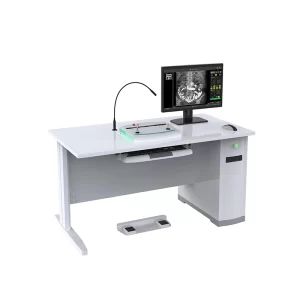
The AR-7900H workstation supports patient registration, work lists, examination records, emergency registration, image storage, report editing, printing, and disc burning. It also includes measurement tools for distance, angle, spine assessment, and cardiothoracic ratio. DICOM printing and network storage support a more practical hospital workflow.
How Does Digital Radiography Help Clinicians Find Abnormalities Earlier?
Digital radiography can support earlier identification of abnormalities and injuries in several ways. On the one hand, static images of the anatomy at a certain point in time are captured. On the other hand, by means of dynamic fluoroscopy, the doctor can watch how a part of the body, or the flow of contrast agents within it, moves. Using both can provide a fuller view when a single image is not enough.
Static Imaging for Common Examinations
Routine digital radiography remains one of the most common hospital imaging methods because it is suitable for many body areas. The AR-7900H supports examinations of the head, chest, lumbar spine, abdomen, and limbs.
A chest image may show changes that call for closer review. A limb or spine image may reveal alignment problems, fractures, or structural changes. The key point is not that one image gives a final diagnosis, but that it can reveal a reason to investigate sooner.
Dynamic Fluoroscopy for Real-Time Observation
Static radiography captures a still image. Fluoroscopy displays a moving image, which can be useful when the doctor needs to observe an area during the examination rather than review only one exposure afterward.
The system supports chest and abdominal fluoroscopy, as well as contrast examinations such as esophagography, pyelography, and salpingography. It can also provide real-time fluoroscopic guidance during foreign-body removal procedures, puncture procedures, and fracture reduction. These imaging capabilities extend the room beyond standard radiography.
Precise and Specialized Imaging for Complex Cases
Some examinations need a targeted point image, an oblique angle, or a wider anatomical view. The system supports precise radiography for lesion observation and point-film positioning, oblique photography for special body parts, standard chest imaging, and whole-spine or lower-extremity stitching.
This matters because early clinical signs are not always located in a convenient position. Flexible imaging gives the operator more ways to capture the area the doctor actually needs to review.
How Does the ARI AR-7900H Support a Broader Imaging Service?
The main strength of the AR-7900H is not one isolated specification. It is the way the functions work together. A hospital can use the combined radiography and fluoroscopy platform for routine imaging, dynamic examinations, contrast studies, special positioning, and image management within one department.
That can be useful for a new hospital, a diagnostic center adding services, or an existing radiology department replacing older equipment. Space is often limited, and buying two separate systems may not be practical. One multifunctional unit can make room planning simpler while giving the department a wider examination range.
The system is also fitted with a 10.4-inch table-side touch screen. Operators can view images and check the source-to-image distance, exposure settings, X-ray field size, tube angle, patient body type, and emergency examination information close to the table. This is a small workflow detail, but it can reduce back-and-forth movement between the patient and the main workstation.
Which Technical Features Matter in Daily Hospital Use?
Specifications should always be connected to real work. A larger detector, for example, is useful because it covers a broader area, not because the number looks impressive on a quotation.
Large Dynamic Flat-Panel Detector
The system has a 427 × 427 mm active imaging area, a 3072 × 3072 pixel matrix, a 139 μm pixel pitch, 16-bit conversion, and an acquisition speed of up to 30 frames per second. The large detector provides a broad field for routine examinations, while the frame rate supports dynamic imaging.
A larger field can also reduce the need to divide some examinations into several small exposures. For a busy department, fewer unnecessary repeats mean less time spent bringing the patient back into position.
High-Frequency Generator and Exposure Control
The AR-7900H uses a 50 kW high-frequency generator with a 500 kHz inverter frequency. The radiography tube voltage range is 40 to 150 kV, and the tube current range is 10 to 630 mA. Continuous and pulsed fluoroscopy settings are also available.
Different body areas and patient sizes need different exposure conditions. A practical system should give the operator enough control to select settings for thin adults, average adults, and larger patients while keeping the examination workflow clear.
Digital Processing and Image Management
The workstation includes zoom, rotation, image inversion, magnification, window-level adjustment, measurement, labeling, report editing, storage, printing, and network configuration. AEC, whole-spine stitching, a dose-area product device, and a high-density grid are listed as standard functions or configurations on the product page.
These tools do not replace the radiologist. They help the clinical team review the image, measure visible structures, keep records organized, and share results through the hospital.
What Should Hospitals Check Before Purchase, Installation, and Service?
Before asking for a quotation, list the examinations your department expects to perform. If you need only basic chest and limb radiography, your priorities may be different from those of a hospital that also handles fluoroscopy, contrast studies, trauma work, full-spine imaging, and lower-extremity stitching.
Room and electrical conditions must also be checked early. The published specification calls for 380V ±38V, 50Hz ±1Hz, and a power capacity of at least 85 kVA. The table has a 200 kg load capacity. These details affect room preparation, power planning, patient access, and installation cost.
You should also ask how the workstation will connect with your current image storage and reporting process. Confirm the standard configuration, optional items, installation responsibilities, staff training, warranty, spare-part availability, and technical response procedure before placing the order.
ARI states that X-ray machines carry a 14-month warranty and that spare parts and product support are available for no less than eight years from delivery. Its service support also covers repair arrangements and replacement parts under the stated warranty conditions.
For a useful project discussion, prepare your hospital type, expected examination range, daily patient volume, room dimensions, local voltage, digital workflow requirements, and target installation date. You can then contact ARI for configuration details, installation questions, training arrangements, and a project quotation.
FAQ
Q1: How Does Digital Radiography Equipment Support Earlier Identification of Abnormalities?
A1: It provides clinicians with timely images of internal structures, helping them identify suspicious changes and determine whether further examination, clinical evaluation, or treatment is needed.
Q2: Can the AR-7900H Perform Both Radiography and Fluoroscopy?
A2: Yes, the system combines digital radiography and digital fluoroscopy in one platform for static imaging, dynamic observation, contrast studies, and selected fluoroscopy-guided procedures.
Q3: Which Body Areas Can Be Examined With the AR-7900H?
A3: It supports imaging of the head, chest, lumbar spine, abdomen, limbs, whole spine, and lower extremities, together with several fluoroscopy and contrast examination applications.
Q4: What Should a Hospital Prepare Before Installing the System?
A4: You should confirm room dimensions, radiation protection requirements, power supply, equipment access, patient flow, workstation networking, operator training, and local installation rules.
Q5: Does the AR-7900H Provide an Automatic Diagnosis?
A5: No, the system produces and processes medical images, while qualified clinicians interpret those images together with the patient’s symptoms, history, examination findings, and other test results.